Diagram Of The Integumentary System
Integumentary system functions and diseases. Arrector pili muscle.
 All Classes In School Home
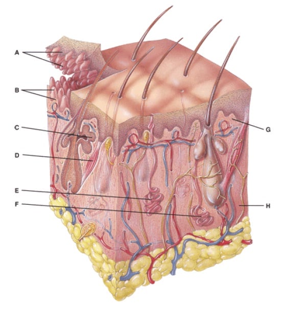
The integumentary system consists of the skin and accessory structures such as hair nails and glands.
Diagram of the integumentary system. Contains spiny shaped cells stratum basale. Integumentary system parts the skin. Found in the epidermis.
It may serve to waterproof cushion and protect the deeper tissues excrete wastes. Diagram of the skin and hair includes sweat gland the stratum granulosum. Learn this topic now at kenhub.
Besides the skin it comprises the hair and nails as well which are appendages of the skin. A fiber protein that is the main component of hair skin and nails. The integumentary system has a variety of functions.
Diagram of the human integumentary system infographic the skin is the largest organ of the body and helps protect it from the environment. Acute or chronic inflammatory skin disease. The deepest layer in the epidermis.
Integumentary system part i. Integumentary system health science 1. Found in the epidermis.
Epidermis 5 layers made up of epithelial tissue only. Structure and layers of the skin. Skeletal system diagrams 11 disorder of the sebaceous glands.
The integumentary system is recognizable to most people because it covers the outside of the body and is easily observed. The deeper subcutaneous stratum on the other hand is made up of connective tissue and fatty substances. The integumentary system is the organ system that protects the body from damage comprising the skin and its appendages including hair scales and nails.
The integumentary system is the set of organs that forms the external covering of the body and protects it from many threats such as infection desiccation abrasion chemical assault and radiation damage. Contains a continuous layer of cells. Sebum plugs pores areas fill contagious fungal infection common in sports lockers.
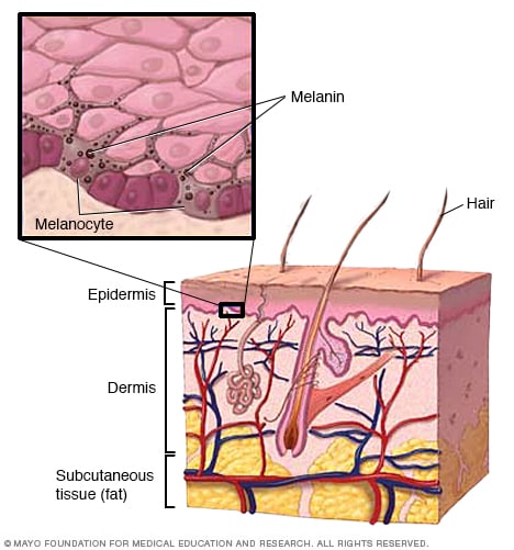
This article defines the integumentary system and discusses its anatomy and function together with clinical aspects. Contains cells that die and move to the surface. Melanocytes are the epidermal cells and are responsible for the synthesis of melanin pigment which in turn renders coloration to the skin.
Hardens and waterproofs the skin. Dermis 2 layers contains connective tissue vessels etc. This article describes the parts of the skin dermis epidermis etc their functions and clinical significance.
The integumentary system or skin is the largest organ in the body.
2015 A P Integumentary Handout
 14 Best Integumentary Images Skin Anatomy Human Anatomy Education
 The Parts Of The Integumentary System Integumentary System Parts And
![]() Diagram Pictures Integumentary System Anatomy Kenhub
Diagram Pictures Integumentary System Anatomy Kenhub
 Integumentary System 2nd Period Swimming Group 6
 Integumentary System Diagram Worksheet Anatomy Pinterest
 Anatomy And Physiology Questions The Integumentary System
 Show Pictures Integumentary System This Skin Diagram Clearly Shows
 Vitiligo Symptoms And Causes Mayo Clinic
 Blank Worksheet System Coloring Diagram Integumentary Page
 Integumentary System 3rd Period Group 2 Basketball
 Hair Diagram Integumentary System

 Human Anatomy Integumentary System Diagrams By Julie Ridge Designs
 Show Pictures Integumentary System This Skin Diagram Clearly Shows

 Game Statistics Skin Diagram Integumentary System Purposegames
Human Anatomy

 Fox Nervous System Diagram Schematic Diagram
 Intugementary System
 Integumentary Anatomy Integumentary System Diagram Integumentary
 Show Pictures Integumentary System This Skin Diagram Clearly Shows
 Diagram Integumentary System Hair Falling
0 Response to "Diagram Of The Integumentary System"
Post a Comment