In The Diagram Of Skin Shown Below Where Is The Apocrine Sweat Gland
Which structure in the figure produces a protein that helps protect the skin and underlying tissues from heat. Contains more sweat glands than thin skin.
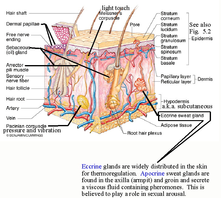
The hypodermis consists primarily of loose connective tissue and lobules of fat.

In the diagram of skin shown below where is the apocrine sweat gland. It is often referred to as subcutaneous tissue though this is a less precise and anatomically inaccurate term. In the diagram of skin shown below where is the reticular region of the dermis. An apocrine sweat gland ˈæpəkrən ˌkraɪn ˌkriːn.
A layer of tissue lies immediately below the dermis of vertebrate skin. Does not contain hair follicles. Which of the following terms refers to paleness of the skin such as seen in patients with shock or anemia.
Which of the following terms refers to paleness of. The epidermal ridge pattern is in part genetically determined and is unique for each individual. This pin was discovered by summer ekelund.
Medium learning objective 1. The ducts of sweat glands open on the tops of the epidermal ridges as sweat pores the sweat and ridges form fingerprints or footprints on touching a smooth object. 51 describe the composition of the epidermis and dermis.
In the diagram of the skin shown below where is the arrector pili muscle. From greek apo away and krinein to separate is composed of a coiled secretory portion located at the junction of the dermis and subcutaneous fat from which a straight portion inserts and secretes into the infundibular portion of the hair follicle. It is used mainly for fat storage.
A e b f c g d h e a ans. Discover and save your own pins on pinterest. Normally the ridge pattern does not change during life.
Found in the palms soles of the feet and fingertips.
The Human Eccrine Sweat Gland Structure Function And Disorders
 Skin Junqueira S Basic Histology Text And Atlas 15e
 The Integumentary System Lesson 0384 Tqa Explorer
 Skin Junqueira S Basic Histology 14e Accessmedicine Mcgraw
 Sweat Gland And Sebaceous Gland Images Stock Photos Vectors
 Histological Analysis Of Human Eccrine Sweat Glands Monolayer Cells
 18 This Is Fine Nonpigmented Hair That Covers The Body Of The Fetus
 Midterm Anatomy Physiology 215 With Rush At Salish Kootenai
 Pdf Defining Key Genes Regulating Morphogenesis Of Apocrine Sweat
 Skin Junqueira S Basic Histology 14e Accessmedicine Mcgraw
 Monitoring Of Sweat Secretion From Eccrine Sweat Glands Using
 Epidermis Wikipedia

07c Investigating The Skin
 Ch 5 Quizlet 1 Pdf Anatomy And Physiology Unit 5 Quiz
 The Integumentary System Lesson 0384 Tqa Explorer
 Localization Of Nestin Positive Cells In Human Sweat Glands Of
 The Integumentary System Lesson 0384 Tqa Explorer
 Mesenchymal Stem Cells For Sweat Gland Regeneration After Burns
 Test Chpater 5 Plqs Quizlet
 Midterm Flashcards Quizlet
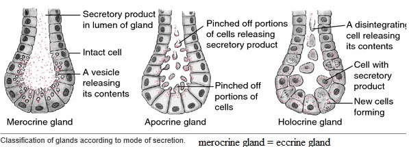
Sweat Or Sudoriferous Glands Include Apocrine And Merocrine Glands
 The Microfluidics Of The Eccrine Sweat Gland Including Biomarker
0 Response to "In The Diagram Of Skin Shown Below Where Is The Apocrine Sweat Gland"
Post a Comment