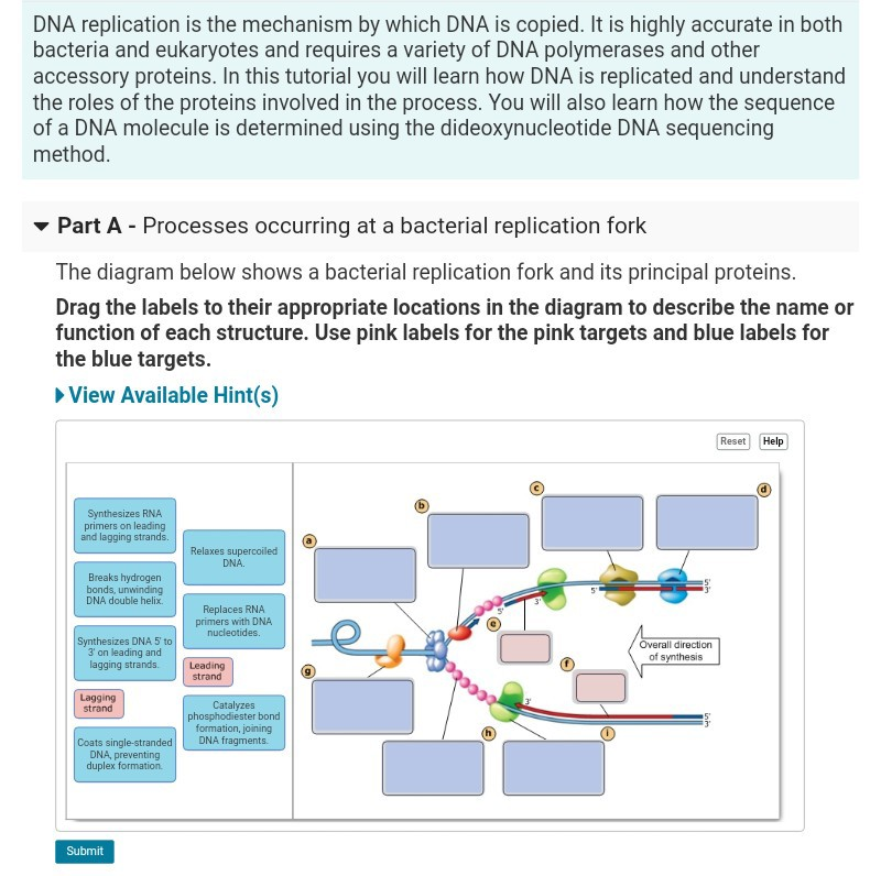
The Diagram Below Shows A Bacterial Replication Fork And Its Principal Proteins
Drag the labels to their appropriate locations in the diagram to describe the name or function of each structure. Use pink labels for the pink targets and blue labels for the blue targets.
Recq And Fe S Helicases Have Unique Roles In Dna Metabolism Dictated
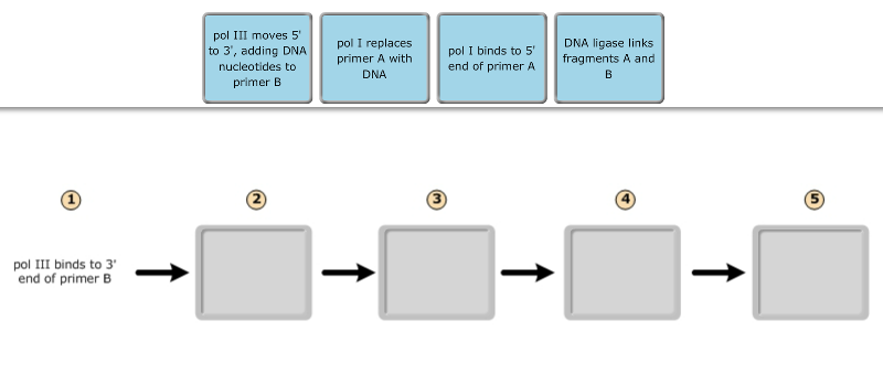
Fragment a is the most recently synthesized okazaki fragment.

The diagram below shows a bacterial replication fork and its principal proteins. Mastering biology exam 3. Part b processes occurring at a bacterial replication fork the diagram below shows a bacterial replication fork and its principal proteins. During a second cycle of replication all four strands in the two dna molecules will serve as templates resulting in four molecules eight strands of dna.
Use pink labels for the pink targets and blue labels for the blue targets. Correct answer below shows a bacterial replication fork and its principal proteinsdrag the labels to their appropriate locations in the diagram to describe the name or function of each structure. Use pink labels for the pink targets and blue labels for the blue targets.
In contrast to the leading strand the lagging strand is synthesized as a series of segments called okazaki fragments. The diagram below shows a bacterial replication fork and its principal proteins. The diagram below shows a replication fork with the two parental dna strands labeled at their 3 and 5 ends.
Part c synthesis of the lagging strand. Drag the labels to their appropriate locations in the diagram to describe the name or function of each structure. Part a the mechanism of dna replication the diagram.
The diagram below shows a bacterial replication fork and its principal proteins. Drag the labels to their appropriate locations in the diagram to describe the name or function of each structure. The diagram below shows a bacterial replication fork and its principal proteins.
Use pink labels for the pink targets and blue labels for the blue targets. Drag the labels to their appropriate locations in the diagram to describe the name or function of each structure. The newly synthesized dna strands are not shown but the polymerase on each parental strand is shown labeled 1 and 2.
The diagram below shows a bacterial replication fork and its principal proteins. Part b processes occurring at a bacterial replication fork the diagram below shows a bacterial replication fork and its principal proteins. Drag the labels to their appropriate locations in the diagram to describe the name or function of each structure.
The diagram below shows a bacterial replication fork and its principal proteins. The diagram below illustrates a lagging strand with the replication fork off screen to the right.
 Part A The Mechanism Of Dna Replication The Diagram Below Shows A Double
 Proposal For A Minimal Dna Auto Replicative System Intechopen
Heterogeneity Of Spontaneous Dna Replication Errors In Single
 Dna Replication Leading Vs Lagging Strand Youtube
 The Diagram Below Shows A Bacterial Replic Clutch Prep
 Solved Dna Replication Is The Mechanism By Which Dna Is C
 Solved Biology Question Chegg Com
 Proposal For A Minimal Dna Auto Replicative System Intechopen
Dna Polymerase Dna Encyclopedia
 The Diagram Below Shows A Bacterial Replic Clutch Prep
 The Importance Of Repairing Stalled Replication Forks Nature
 Dna Replication Fork Definition Overview Video Lesson
 The Diagram Below Shows A Bacterial Replic Clutch Prep
 Mastering Biology Chapter 16 Rhs Homework
 Chaperoning Hmga2 Protein Protects Stalled Replication Forks In Stem
Dna Polymerase Iv Primarily Operates Outside Of Dna Replication
 Transcription Leads To Pervasive Replisome Instability In Bacteria
Station F Dna Replication F1 F6 Fill In The Blanks
 Exam 3 Chs 5 Dna Structure And Replication Machinery 16 The
 Exam 3 Chs 5 Dna Structure And Replication Machinery 16 The
 The Diagram Below Shows A Bacterial Replic Clutch Prep
0 Response to "The Diagram Below Shows A Bacterial Replication Fork And Its Principal Proteins"
Post a Comment