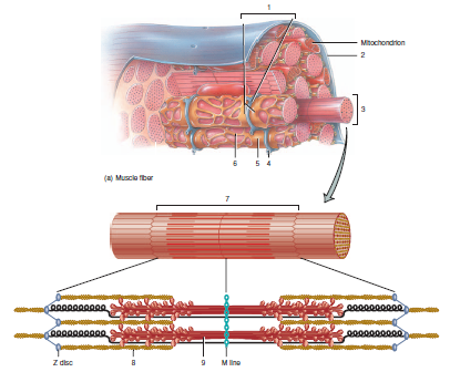
Drag The Labels Onto The Diagram To Identify Structural Features Associated With Skeletal Muscle
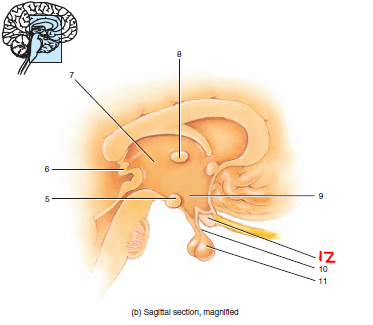
Maintain posture maintain body temperature guard body entrances. Numbers to the left identify the spinal nerves and indicate where the nerve roots leave the vertebral canal.
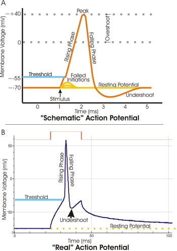 Threshold Potential Wikipedia
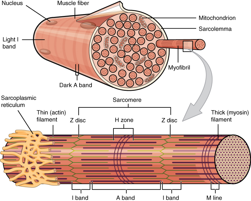
Muscle covered with epimysium made up of fascicles bundle of cells covered in perimysium made up of muscle fibers cells covered in endomysium myofibrils made of molecular motors make up each muscle fiber and sarcomeres are contained in each myofibril blood vessels line in between the connective tissue coverings in between.

Drag the labels onto the diagram to identify structural features associated with skeletal muscle. Drag the labels onto the diagram to identify structural features associated with skeletal muscle. Show transcribed image text drag the labels onto the diagram to identify structural features associated with skeletal muscle. Golgi apparatus renewal or modification of the cell membrane is a function of the lysosomes each of the following is an example of a non membranous organelle except digestion.
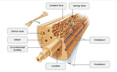
Mastering ap exam 1. Skeletal smooth cardiac all of these muscle types have about the same effect on the bodys heat production. The structure of bone tissue suits the function.
Study ap chapter 6 bones and skeletal tissues flashcards taken from chapter 6 of the book human anatomy physiology. Drag the appropriate labels to their respective targets. When an antigen is bound to a class ii mhc protein it can activate a cell.
This suggests that week four assignment three 8 drag the labels onto the diagram to identify the components of a model cell. Part a drag the labels onto the diagram to identify structural features associated with skeletal muscle. Part a drag the labels onto the diagram to identify structural features associated with a skeletal muscle fiber submit request answer show transcribed image text the labels onto the diagram to identify structural features associated with a skeletal muscle fiber.
The structure of a muscle cell can be explained using a diagram labelling muscle filaments myofibrils sarcoplasm cell nuclei nuclei is the plural word for the singular nucleus sarcolemma and the fascicle of which the muscle fibre is part. The structure of muscle fibers is included in courses in human biology and human anatomy and physiolgy. Innervates skeletal muscles of the neck and back.
Features of the spinal cord 45 cm in length passes through the foramen magnum. The spinal cord however. Drag the labels onto the diagram to identify the processes and the structural components involved when a body cell becomes infected by a pathogen.
Which of the following bone tissues is adapted to support weight and withstand tension stress.
 Lab 9 Muscle Physiology
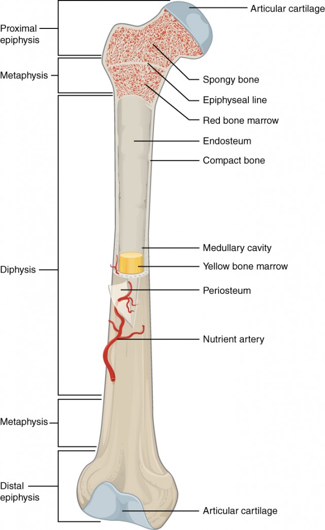 Bone Structure Anatomy And Physiology I
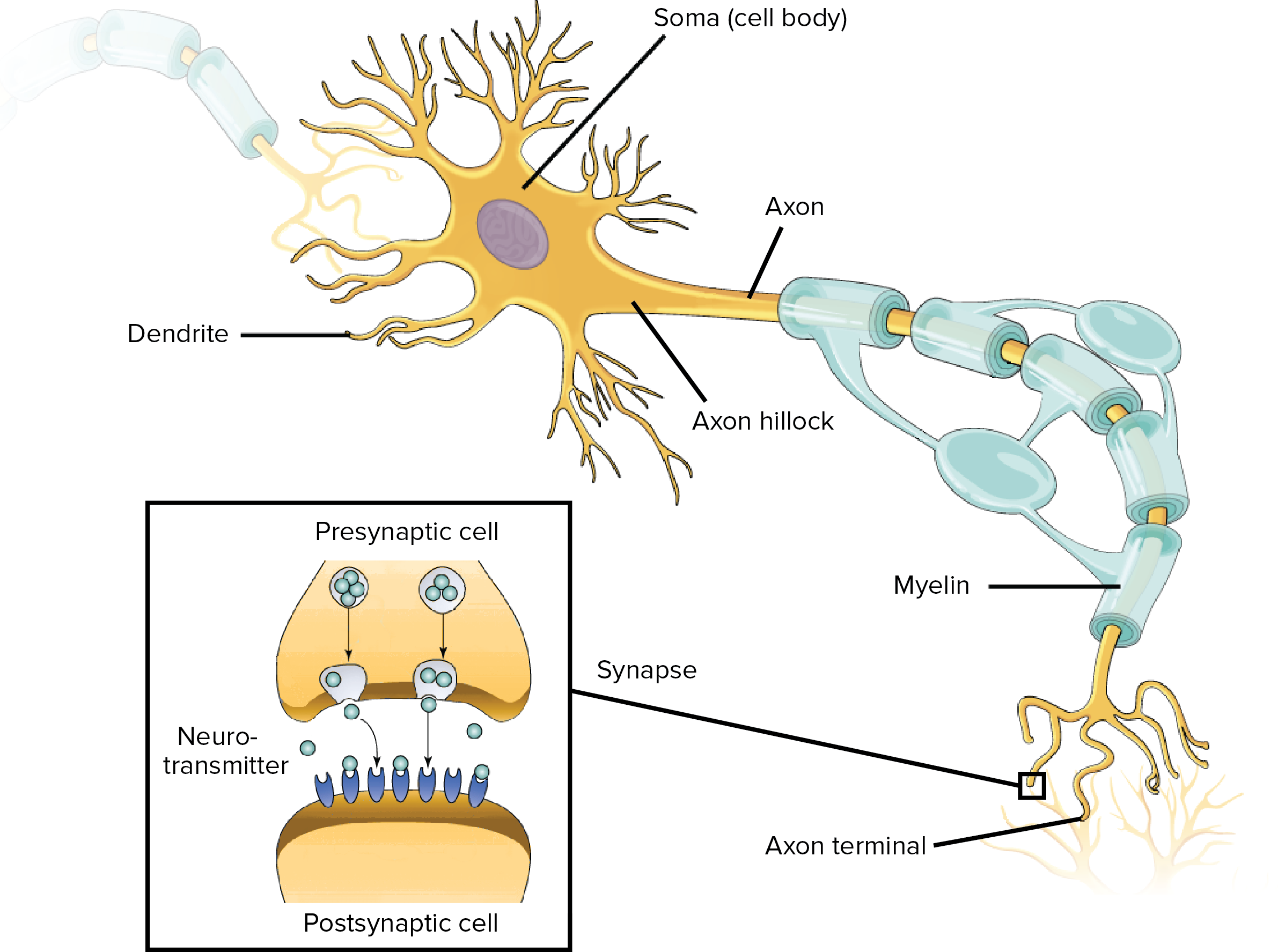 Human Biology Ap Biology Science Khan Academy
Solved Lables Onto The Diagram To Identify Structural Fea
 Solved Art Labeling Activity Figure 15 1 Drag The Labels
 28 Best Kathrin Images On Pinterest Anatomy Physical Therapy And
 Anatomy Exam 2 Flashcards Easy Notecards
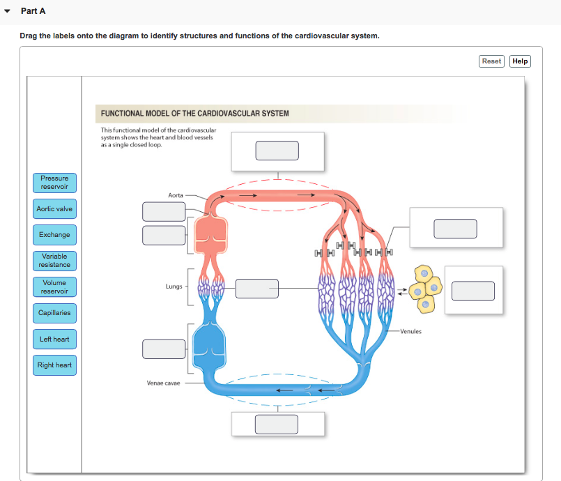
 Heart Structure Anatomy Physiology Wikivet English
The Nervous System The Spinal Cord And Spinal Nerves
Lab 9 Muscle Physiology
Label The Components Of A Knee Jerk Reflex Part A Chegg Com
 10 2 Skeletal Muscle Anatomy And Physiology
Solved The Labels Onto The Diagram To Identify Structural
Solved Drag The Labels Onto The Diagram To Identify Struc
 A P Chapter 6 Bones And Skeletal Tissues Flashcards Easy Notecards
 Answer Correct Chapter Test Chapter 10 Question 4 Endomysium Perimysium
Answer Correct Chapter Test Chapter 10 Question 4 Endomysium Perimysium
 Heart Structure Anatomy Physiology Wikivet English
 Anatomy Exam 2 Flashcards Easy Notecards
 14 5 Sensory And Motor Pathways Anatomy Physiology

0 Response to "Drag The Labels Onto The Diagram To Identify Structural Features Associated With Skeletal Muscle"
Post a Comment