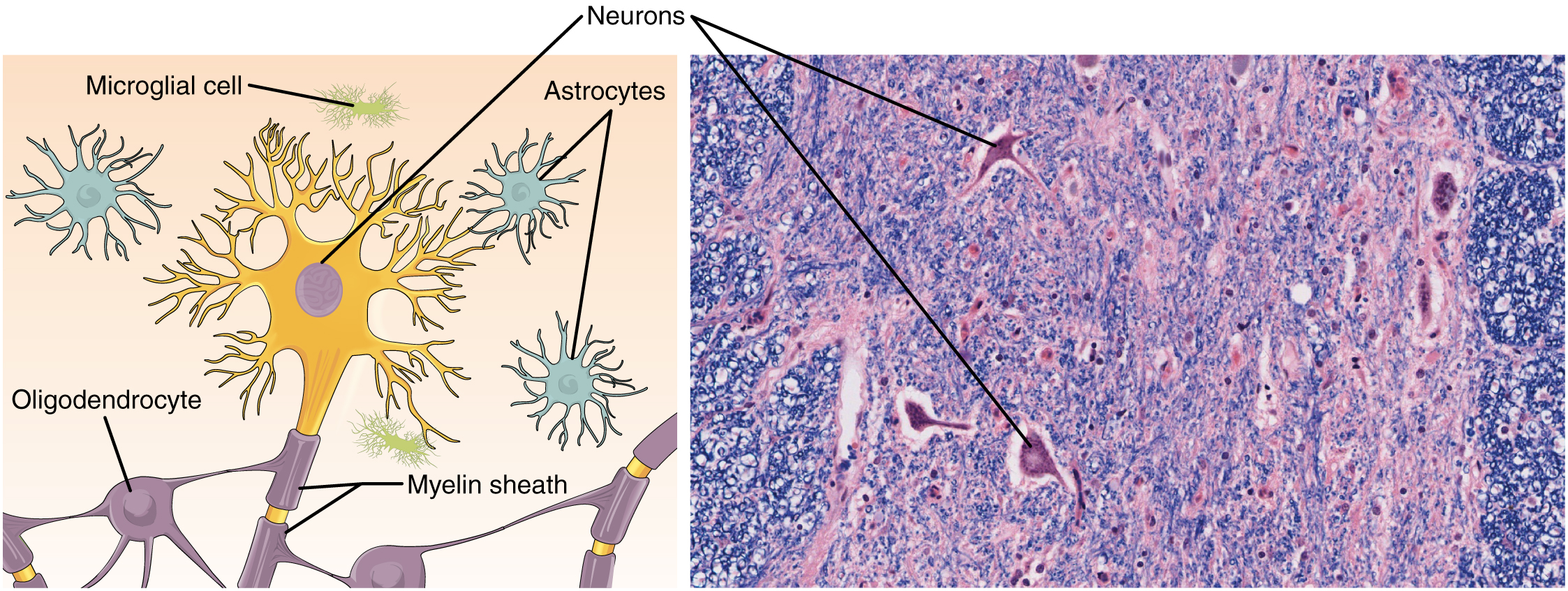
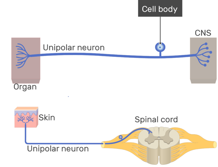
What Specific Type Of Unipolar Neuron Is Shown In The Diagram Labeled B
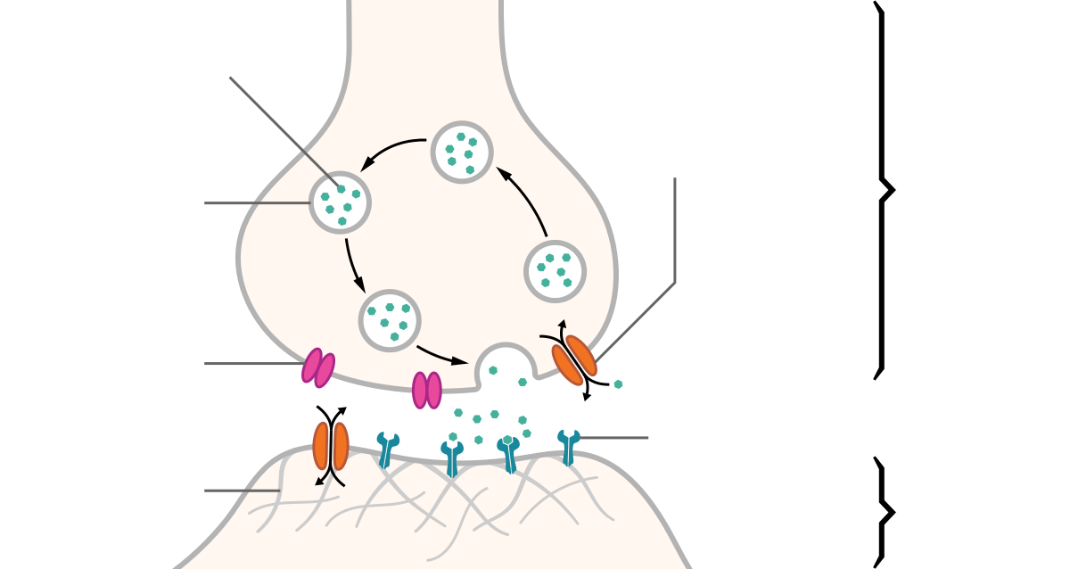
4presyn neuron releases chem messengers called 5synapse of a neuron and a gland is called a neuromusc junct. D parasympathetic postganglionic neuron.

D parasympathetic postganglionic neuron.

What specific type of unipolar neuron is shown in the diagram labeled b. 3synapse is the site where 2 neurons or a neuron an effector communicate. What specific type of unipolar neuron is shown in the diagram labeled b what specific type of unipolar neuron is shown in the diagram labeled b nervous system. A body b axon c dendrite d synapse a.
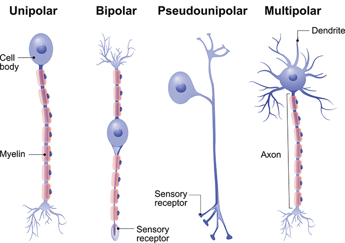
B a merkel disc is a touch receptor that consists of free nerve endings bare dendrites that make contact with merkel cells of the stratum basale of the skin. A somatic motor neuron. B parasympathetic preganglionic neuron.
What specific type of unipolar neuron is shown in the diagram labeled c. A merkel disc b meissner corpuscle c pacinian corpuscle d nociceptor e purkinje cell ans. Easy learning objective 1.
What specific type of unipolar neuron is shown in the diagram labeled b photos. What specific type of unipolar neuron is shown in the diagram labeled b. E sympathetic preganglionic neuron.
E sympathetic preganglionic neuron. A type i cutaneous mechanoreceptor merkel disc b corpuscle of touch meissner corpuscle. 1presyn neuron carries a nerve impulse away from a synapse.
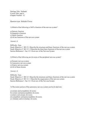
What specific type of unipolar neuron is shown in the diagram labeled b. 122 describe the structures and functions of neurons and neuroglia and distinguish between white matter and gray matter. The structure labeled 3 in the diagram is a a somatic motor neuron.
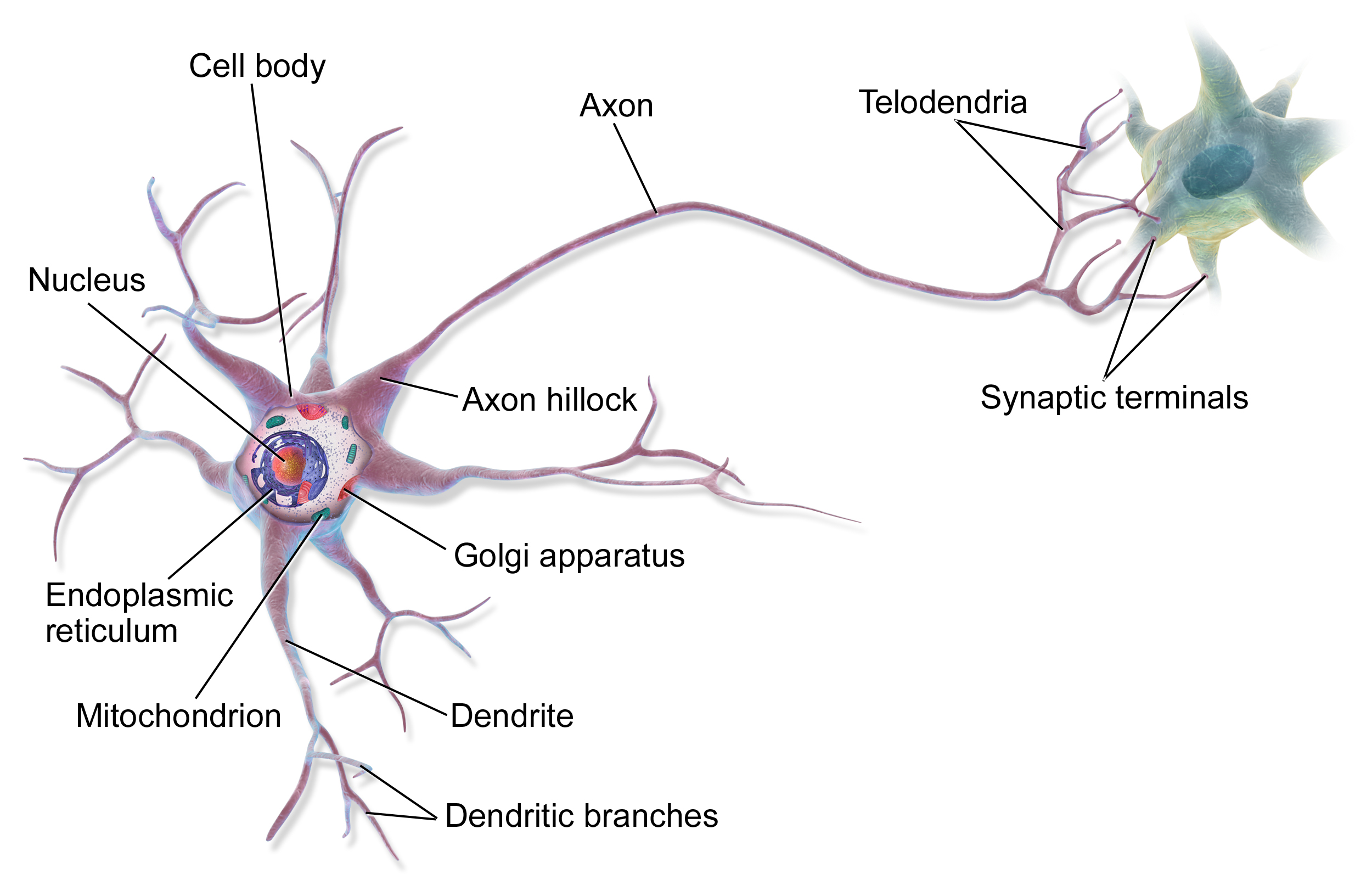
2postsyn neuron carries a nerve impulse toward a synapse. Which part of the neuron is responsible for cell metabolism. C a pacinian corpuscle is a pressure receptor composed of a multilayered connective tissue capsule that encloses a dendrite.
Which labeled cell is located mainly in white matter and helps maintain appropriate chemical environment for generation of nerve impulses. The structure labeled 3 in the diagram is a. Correct answer below a type i cutaneous mechanoreceptor merkel discb corpuscle of touch meissner corpusclec lamellated pacinian corpuscled nociceptore purkinje cell.
B parasympathetic preganglionic neuron. E cerebellum this major portion of the brain is used to monitor movements initiated by the motor areas of the cerebrum. Which of the labeled structures in the diagram is the thalamus.
 Exam 2016 Biol 235 Human Anatomy And Physiology Studocu
 Ch 12 Diagrams For The Final Study Anatomy Physiology 2010
 Unipolar Neuron Structure And Functions
 A A B B C C D D Answer D 55 What Is The Structural Classification Of
 What Specific Type Of Unipolar Neuron Is Shown In The Diagram
 4 5 Nervous Tissue Mediates Perception And Response Anatomy And
 Types Of Neurons Queensland Brain Institute University Of Queensland
 Human Biology Science Khan Academy
Rabconnectin 3a Is Required For The Morphological Maturation Of Gnrh
 Nervous Tissue Anatomy And Physiology I
 Synapse Wikipedia
 Nervous Tissue Anatomy And Physiology I


 Nerve Tissue The Nervous System Junqueira S Basic Histology 14e
 Unipolar Neuron Structure And Functions
 Axon Wikipedia
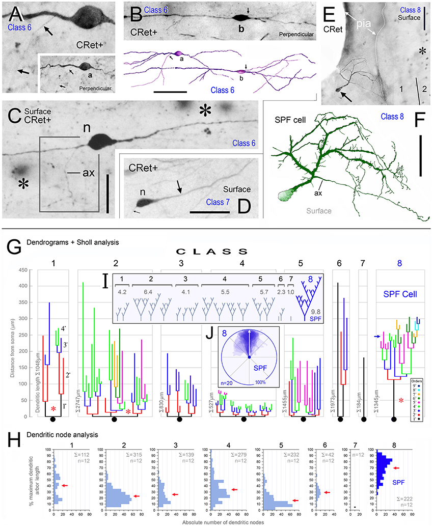 Frontiers Subpial Fan Cell A Class Of Calretinin Neuron In
0 Response to "What Specific Type Of Unipolar Neuron Is Shown In The Diagram Labeled B"
Post a Comment